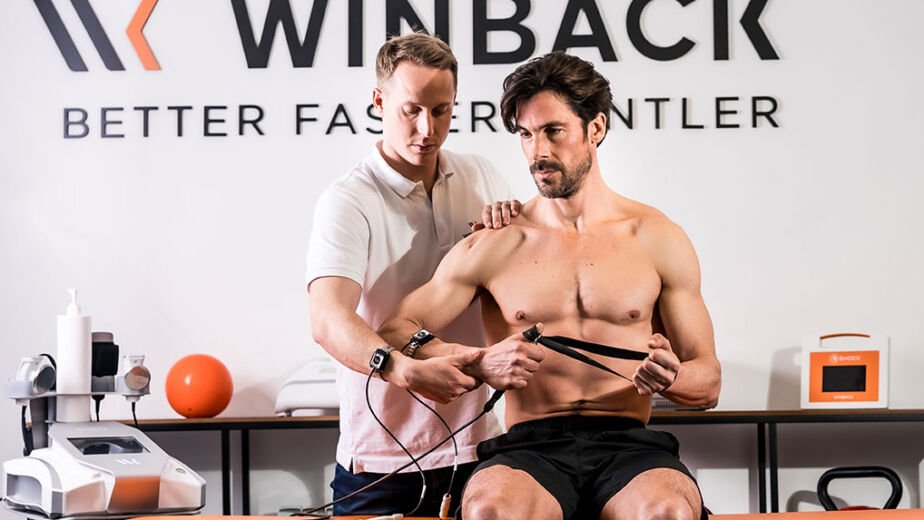
Musculoskeletal injury does more than damage tissue—it alters the way the nervous system drives the muscle. Reflexive inhibition, known as arthrogenic muscle inhibition (AMI), is a well-documented barrier to rehabilitation. Pain, effusion, and altered joint afferents reduce motor neuron excitability, leading to decreased muscle activation, atrophy, and delayed return to function. Traditional rehabilitation often struggles against this invisible brake. Emerging evidence supports the use of Winback TECAR therapy and electrical muscle stimulation (EMS) as complementary strategies to re-establish motor control and accelerate recovery.
The Problem: Injury-Induced Inhibition
Injury-induced inhibition, often referred to as arthrogenic muscle inhibition (AMI), describes the reflexive downregulation of muscle activation that occurs after joint or soft tissue trauma. When an injury produces pain, effusion, or capsular swelling, altered afferent input from mechanoreceptors and nociceptors in the joint is transmitted to the spinal cord and motor cortex. This disrupts normal motor neuron excitability, resulting in decreased voluntary activation of surrounding musculature even when the patient attempts maximal contraction.
The phenomenon is well documented in the quadriceps after ACL injury and reconstruction, where persistent activation deficits of up to 20–40% have been reported despite apparent recovery of strength (Pietrosimone et al., 2015).
Similar effects have been observed in controlled models of knee effusion, where as little as 20–30 mL of intra-articular fluid induces significant reflex inhibition of quadriceps activity (Rice et al., 2014). Beyond the knee, inhibition has also been demonstrated in the hamstrings following strain injuries, particularly at longer muscle lengths where eccentric weakness is most pronounced (Opar et al., 2015).
The peroneals and soleus show comparable reductions in excitability after ankle sprain, contributing to chronic instability (Palmieri-Smith et al., 2019), and patients with chronic rotator cuff tears exhibit abnormal activation patterns in the deltoid consistent with central neuromuscular dysfunction (de Oliveira et al., 2015).
Left unaddressed, AMI delays rehabilitation, accelerates atrophy, disrupts motor coordination, and increases reinjury risk, underscoring the importance of interventions that target both pain/effusion and neuromuscular drive.
The Opportunity: TECAR and EMS in Muscle Re-education
TECAR (Transfer of Energy Capacitive and Resistive) applies high-frequency current that interacts with tissue capacitance and resistance. Its therapeutic benefits are twofold:
- Pain and Effusion Modulation – TECAR increases circulation and lymphatic drainage, reduces nociceptive signaling, and improves viscoelastic properties. By mitigating pain and swelling (two primary AMI drivers) TECAR helps “unlock” motor inhibition.
- Neuromodulation – CET and RET currents act on sensory pathways within the joint, reducing abnormal signaling that drives reflexive inhibition. By modulating these joint afferents and spinal reflex loops, they help restore normal excitability of the motor neurons and improve muscle activation.
Clinical Example: In ACL rehabilitation, TECAR applied to the knee joint reduces pain and effusion, allowing subsequent quadriceps exercises to be performed with less inhibition and greater voluntary activation.
Electrical Muscle Stimulation (EMS)
EMS directly recruits motor units by bypassing impaired voluntary drive. When synchronized with patient effort, EMS reinforces motor patterns at both the peripheral and cortical levels.
- Isometric re-education: EMS-assisted quadriceps sets restore recruitment in inhibited muscles.
- Functional integration: Layering EMS onto dynamic movement promotes motor learning and prevents maladaptive compensation.
Clinical Example: In hamstring strain rehabilitation, EMS applied during lengthened isometric holds enhances neuromuscular drive, addressing the selective eccentric weakness identified post-injury.
Application: TECAR + EMS
The versatility of the Winback TECAR system allows for a multi-modal approach simultaneously. By utilizing the adhesive pads, the provider can now apply EMS current while delivering on the other traditional modes. Providing either a completely hands-free option or semi-hands free.
Knee Effusion and Quadricep Inhibition
This set up shows the option of utilizing double adhesives for the application of EMS on the quadriceps, providing a complete hands-free option to the practitioner. The alternate set up would utilize one adhesive pad, while simultaneously applying other therapeutic modes using the handle to deliver CET or RET and Hi-TENS. While EMS function runs.
Capacitative (CET) Mode allows for superficial circulation and lymphatic drainage, reducing joint swelling, one of the key triggers of quadricep inhibition. Resistive Mode (RET) penetrates deeper to influence joint capsule and periarticular tissues, normalizing afferent feedback from mechanoreceptors disrupted by effusion. Hi-TENS provides analgesia through spinal gating, reducing nociceptive input that compounds inhibition. Once inhibitory environment is quieted, EMS re-educates the quadriceps by directly recruiting motor units pairing EMS with volitional effort trains cortical drive to overcome AMI.
Together, these tools bridge the gap between structural healing and functional restoration. They accelerate the timeline by addressing both sides of AMI: the afferent-driven inhibition and the efferent recruitment deficit.
Conclusion
AMI is a hidden barrier to musculoskeletal rehabilitation. Standard exercise approaches are often insufficient to overcome the reflexive neural inhibition following injury. By combining TECAR’s capacity to normalize the inhibitory environment with EMS’s ability to directly re-educate muscle activation, clinicians can accelerate recovery, reduce recurrence, and restore optimal neuromuscular function.
References
- Hopkins JT, Ingersoll CD. Arthrogenic muscle inhibition: A limiting factor in joint rehabilitation. J Sport Rehabil. 2000;9(2):135–159.
- Hart JM, Pietrosimone B, Hertel J, Ingersoll CD. Quadriceps activation following knee injuries: A systematic review. J Athl Train. 2010;45(1):87–97.
- Spencer JD, Hayes KC, Alexander IJ. Knee joint effusion and quadriceps reflex inhibition in man. Arch Phys Med Rehabil. 1984;65(4):171–177.
- Rice DA, McNair PJ. Experimental knee pain produces immediate quadriceps inhibition: A central nervous system response. Med Sci Sports Exerc. 2010;42(3):623–630.
- Opar DA, Williams MD, Shield AJ. Hamstring strain injuries: Factors that lead to injury and re-injury. Sports Med. 2015;45(1):23–41.
- Palmieri-Smith RM, Hopkins JT, Brown TN. Peroneal activation deficits in persons with functional ankle instability. Am J Sports Med. 2008;36(2):216–223.
- de Oliveira VM, Pitangui AC, dos Santos Neto LD, Araújo RC. Neuromuscular control in chronic rotator cuff tear: Evidence of central dysfunction. Clin Biomech. 2015;30(8):879–885.


