
Authors: Mercedes Furió-Valverde¹²³, Teresa E. Fernández-Pardo¹⁴⁵, Goreti Font Mas⁶, Marta Nieves-Martín¹, Esther Delgado-Pérez¹²
¹ Department of Physiotherapy, Faculty of Medicine, Health and Sports, Universidad Europea de Madrid, Madrid, Spain.
² Woman & Health Research Group, Department of Physiotherapy, Universidad Europea de Madrid, Madrid, Spain.
³ Department of Physiotherapy, Hospital Ruber Internacional, Madrid, Spain.
⁴ Escuela de Doctorado, Universidad Autónoma de Madrid, Madrid, Spain.
⁵ Physiotherapy Department, Ramón y Cajal University Hospital, Madrid, Spain.
⁶ Gabinet Mèdic Girona, Girona, Spain.
Treatment Location: Hospital Ruber Internacional, Madrid, Spain
Key Words: Open wound; traumatic wound; wound healing; granulation tissue; radiofrequency; TECAR; CET; RET; physiotherapy; skin graft; tissue repair; perilesional; fibroblast; angiogenesis
Summary (Abstract)
A previously healthy adult sustained a high-energy motorcycle accident resulting in an open fracture of the left ankle with approximately 7×9 cm of soft tissue loss. Following emergency debridement, six days in the ICU, and approximately one month of hospitalization, the wound remained unsuitable for skin grafting at discharge (Day 29) due to insufficient granulation tissue. With multidisciplinary agreement between the trauma and plastic surgeons, perilesional TECAR therapy (CET and RET) was initiated immediately after discharge and delivered across 15 sessions over 30 days (Days 29–62). A stage-adapted protocol was used: mild thermal CET/RET during the proliferative phase (Sessions 1–9), transitioning to athermal RET only after plastic surgeon reassessment confirmed adequate granulation tissue at Session 9. TECAR was applied exclusively to viable perilesional tissues throughout; the wound bed was never directly treated. Serial photographic documentation demonstrated progressive granulation tissue development, wound area reduction, and advancing epithelialization. The wound reached graft-eligible status within the protocol window. No adverse events were documented.
Key take-away lessons:
- Perilesional TECAR (CET/RET) can be safely integrated into multidisciplinary wound management from the day of hospital discharge, supporting the biological environment of the proliferative phase without adverse events — provided it is never applied directly to the open wound bed.
- A stage-adapted protocol is essential: mild thermal CET/RET during active proliferation to support perilesional perfusion and cellular metabolism, transitioning to athermal RET once the wound moves into remodeling.
- Multidisciplinary coordination (trauma surgeon, plastic surgeon, physiotherapist) is the prerequisite for safe early physiotherapy in complex open wounds. Protocol changes must be driven by specialist reassessment, not by the physiotherapy team acting independently.
Introduction
Extensive traumatic wounds involving loss of skin and underlying soft tissue represent a major challenge for trauma, reconstructive surgery, and rehabilitation teams. When tissue loss is extensive, granulation may progress slowly, with the wound remaining biologically active in the proliferative phase for several weeks — delaying definitive skin grafting and increasing management complexity [1–3]. During this phase, fibroblast activity, angiogenesis, and re-epithelialization are the primary biological targets, and local perfusion and oxygen availability are relevant determinants of granulation tissue quality [1].
Capacitive-resistive energy transfer (TECAR) therapies deliver radiofrequency currents through tissues using capacitive (CET) and resistive (RET) configurations. Experimental work has shown that radiofrequency stimulation can promote fibroblast and keratinocyte proliferation and migration, and can modify proteins involved in cell adhesion and wound closure [6], providing mechanistic plausibility for its use as an adjunct during tissue repair. Most physiotherapy approaches to wound management are introduced after wound closure; far less is known about their role during active repair of a large open wound prior to skin grafting, where close medical supervision, strict avoidance of the wound bed, and continuous protocol adaptation are required.
Patient Information / Diagnosis
Demographics: Previously healthy adult; sex and age not disclosed
Mechanism of Injury: High-speed motorcycle accident
Chief Complaint: Open fracture of the left ankle with extensive soft tissue loss (~7×9 cm defect post-debridement). Wound biologically unsuitable for skin grafting at hospital discharge (Day 29).
Diagnosis: Complex post-traumatic open wound of the left ankle with prolonged proliferative phase and delayed granulation tissue formation.
Relevant Medical / Family History: No relevant prior medical history.
Prior Interventions: Emergency surgical debridement (Day 0); 6 days ICU; approximately one month inpatient wound management including negative pressure wound therapy (NPWT). NPWT reduced wound dimensions from 9×7 cm (post-debridement) to 8×5.8 cm by Day 29. Wound not yet graft-eligible at discharge — granulation tissue still insufficient.
Clinical Findings
Objective — Baseline (Day 29, Session 1)
- Open wound defect at Session 1 (Day 29): 8×5.8 cm (reduced from 9×7 cm post-debridement by NPWT during hospitalization).
- Wound bed: granulation tissue absent or insufficient to support skin graft survival.
- Perilesional tissue: viable; suitable for TECAR application.
- Pain at baseline: 6/10 (VAS) — reduced to 3/10 immediately following Session 4 treatment.
- Primary outcome measure: serial wound measurement and clinical photography at Sessions 1, 4, 6, 9, 12, and 15.
Treatment Timeline and Intervention
Timeline
| Phase | Timepoint | Clinical Events & Intervention |
| Trauma & Emergency Surgery | Day 0 | High-speed motorcycle accident; open fracture left ankle. Post-debridement soft tissue defect: 9×7 cm. Emergency surgical debridement performed. |
| ICU & Hospitalization (NPWT) | Days 1–28 | 6 days ICU; continued inpatient wound management including negative pressure wound therapy (NPWT). NPWT reduced wound dimensions to 8×5.8 cm by Day 29. Wound still unsuitable for skin grafting — granulation tissue insufficient. |
| Physiotherapy Initiated | Day 29 (Session 1) | Hospital discharge. Multidisciplinary agreement (trauma + plastic surgeon) to initiate perilesional CET/RET protocol. Wound: 8×5.8 cm. |
| Proliferative Phase — Thermal Protocol | Sessions 1–9 (Days 29–48) | Mild thermal CET/RET protocol maintained. Progressive granulation tissue formation documented at Sessions 1, 4, 6, and 9. By Session 4 (Day 36): wound 7×4 cm, VAS pain 6→3 post-treatment. By Session 6 (Day 42): complete wound bed coverage with healthy granulation tissue. |
| Phase Transition | Day 48 (Session 9) | Plastic surgeon reassessment confirms satisfactory granulation tissue maturation. Wound transitions from proliferative to remodeling phase. Topical corticosteroid prescribed. Protocol modified: thermal stimulation discontinued; athermal RET only for remaining sessions. |
| Remodeling Phase — Athermal Protocol | Sessions 10–15 (Days 48–59) | Athermal RET mode only. Perilesional application maintained throughout; wound bed never directly treated. Progressive epithelialization from wound margins documented at Session 12 (Day 54). Advanced epithelialization and wound bed maturation confirmed at Session 15 (Day 59). |
Therapeutic Intervention
Type of intervention: Stage-adapted perilesional TECAR therapy (CET and RET) delivered using the Winback BACK device (Winback, France). TECAR was applied exclusively to viable perilesional tissues and never directly to the open wound bed throughout all 15 sessions.
Phase 1 — Mild Thermal Protocol (Sessions 1–9, Days 29–50):
- Stage 1 — 10 min low-intensity CET: neutral plate over gastrocnemius. Prepare surrounding tissues; support cellular metabolic activity.
- Stage 2 — 10 min continuous CET (mild thermal): promote controlled local vasodilation and improve perilesional perfusion during the active proliferative phase.
- Stage 3 — 10 min RET: adhesive return plate over gastrocnemius; neutral plate on plantar foot. Treat deeper perilesional structures while maintaining complete wound bed avoidance.
Phase 2 — Athermal Protocol (Sessions 10–15, Days 51–62):
- Thermal stimulation discontinued following plastic surgeon reassessment confirming satisfactory granulation tissue.
- All remaining sessions: athermal RET only — supporting tissue remodeling without unnecessary thermal stimulation once the granulation bed was established.
Session Frequency / Duration: ~5 sessions/week; approximately 30 minutes per session
Adverse Events: None. No skin reactions, wound deterioration, or TECAR-related adverse events across all 15 sessions.
Results
Wound progression across the 15-session protocol (Table 1):
| Time Point | Day | Session | Wound Size | Clinical Findings |
| Post-debridement | 0 | — | 9×7 cm | Extensive post-traumatic soft tissue defect immediately after surgical debridement. |
| Hospital discharge (after NPWT) | 29 | Before Session 1 | 8×5.8 cm | NPWT during hospitalization reduced wound dimensions. Wound bed still unsuitable for skin grafting — granulation tissue insufficient. Perilesional TECAR protocol initiated at discharge. |
| Session 4 | 36 | 4 | 7×4 cm | Progressive wound contraction; marked reduction in depth from granulation tissue filling the defect. Patient reported mild pruritus. Pain: 6/10 → 3/10 (VAS) immediately after treatment. |
| Session 6 | 42 | 6 | — | Complete coverage of wound bed with healthy, well-vascularized granulation tissue. No exposed deep tissues. Mild pruritus persisted, consistent with active healing. |
| Session 9 | 48 | 9 | — | Continued wound contraction and granulation tissue maturation. Plastic surgeon reassessment: topical corticosteroid prescribed; protocol transitioned from mild thermal to athermal RET. |
| Session 12 | 54 | 12 | — | Progressive epithelialization from wound margins; continued reduction in wound area. |
| End of treatment | 59 | 15 | — | Advanced epithelialization and satisfactory wound bed maturation at completion of physiotherapy programme, allowing continuation of planned reconstructive management. |
Wound dimensions decreased from 9×7 cm at debridement to 8×5.8 cm at Day 29 (after NPWT) and 7×4 cm by Session 4 (Day 36). By Session 6, complete wound bed coverage with healthy granulation tissue was achieved. Pain reduced from 6/10 to 3/10 (VAS) immediately following Session 4. No adverse events associated with TECAR were documented across all 15 sessions.
Clinical photographs from the personal archive of Mercedes Furió-Valverde (Days 0, 29, 36, 42, 48, 54, 59). © Mercedes Furió-Valverde. All rights reserved — not for reproduction without prior written permission.
Discussion
This case demonstrates the clinical feasibility of integrating early perilesional TECAR therapy into the multidisciplinary management of a large post-traumatic open wound — from the day of hospital discharge through to graft-eligible wound status — without adverse events. The protocol achieved its primary objective: development of a well-vascularized granulation tissue bed within a 30-day window. The initial mild thermal CET/RET protocol was selected to support perilesional vasodilation and cellular metabolic activity during the prolonged proliferative phase, a rationale grounded in experimental evidence that radiofrequency currents can promote fibroblast and keratinocyte proliferation and migration [6]. However, blood flow and tissue oxygenation were not directly measured, and the relationship between mild thermal radiofrequency, perfusion, and wound healing remains to be established through controlled trials.
The transition to athermal RET at Session 9 — driven by plastic surgeon reassessment, not physiotherapy protocol alone — reflects the central clinical principle of this case: the therapeutic objective must shift when the wound shifts. Once adequate granulation tissue was confirmed and the wound entered the remodeling phase, continued thermal stimulation was neither necessary nor appropriate. Athermal RET continued to deliver bioelectrical tissue-level effects without it.
Limitations include the single-patient design (causality cannot be established), multiple concurrent wound care interventions that prevent isolation of TECAR’s independent contribution, and the absence of planimetric wound measurement. Future prospective case series should incorporate standardized wound area quantification, tissue oxygenation assessment, and follow-up through skin grafting and post-graft recovery.
References
- Cambronero Ulate P, et al. Fisiopatología de la cicatrización patológica. Rev Med Sinergia. 2022;7(5):e820.
- Lee CC, et al. An updated review of the immunological mechanisms of keloid scars. Front Immunol. 2023;14:1117630.
- Kim HJ, Kim YH. Comprehensive insights into keloid pathogenesis and advanced therapeutic strategies. Int J Mol Sci. 2024;25(16):8776.
- Putri IL, et al. The efficacy of topical oxygen therapy for wound healing: a meta-analysis. Int Wound J. 2024;21(7):e14960.
- Nagarsheth K, et al. Systematic review of the effects of topical oxygen therapy on wound healing. 2024.
- Hernández-Bule ML, et al. In vitro stimulation with radiofrequency currents promotes proliferation and migration in human keratinocytes and fibroblasts. Electromagn Biol Med. 2021;40(3):338–352.
Informed Consent
Patient provided informed consent for treatment and for the de-identified use of clinical data, photographs, and outcome measures in this case report. Documentation available on request.
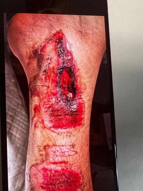
Figure 1
Day 0 – Initial traumatic injury
(before surgical debridement)
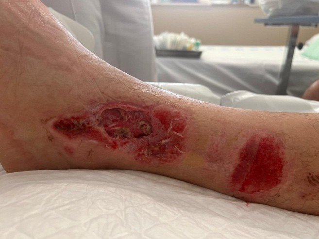
Figure 2
Day 29 – Session 1
Immediately after hospital discharge
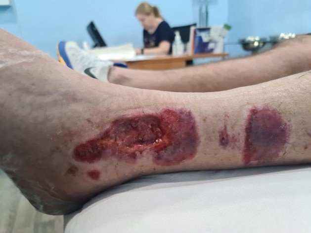
Figure 3
Day 36 – Session 4
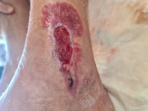
Figure 4
Day 42 – Session 6
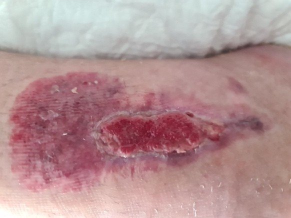
Figure 5
Day 50 – Session 9
Final session using mild thermal protocol protocol following reassessment
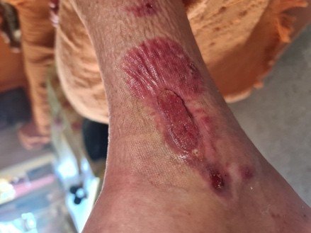
Figure 6
Day 51 – Session 12
Athermal resistive
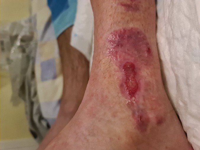
Figure 7
Day 62 – Session 15
End of physiotherapy protocol